5 Chairside Protocols for Crisp Subgingival STL Margin Capture
30-SECOND CLINICAL TAKEAWAYS
6 min read
- Double-cord deflection (#000 + #0) paired with gentle air-drying eliminates subgingival mesh bridging in CAD.
- Color PLY/OBJ exports dramatically improve equigingival finish-line visibility over monochrome STL.
- Upright bilateral buccal bite scans prevent virtual articulator high spots at delivery.
1. Why Optical Line-of-Sight Determines Marginal Fit
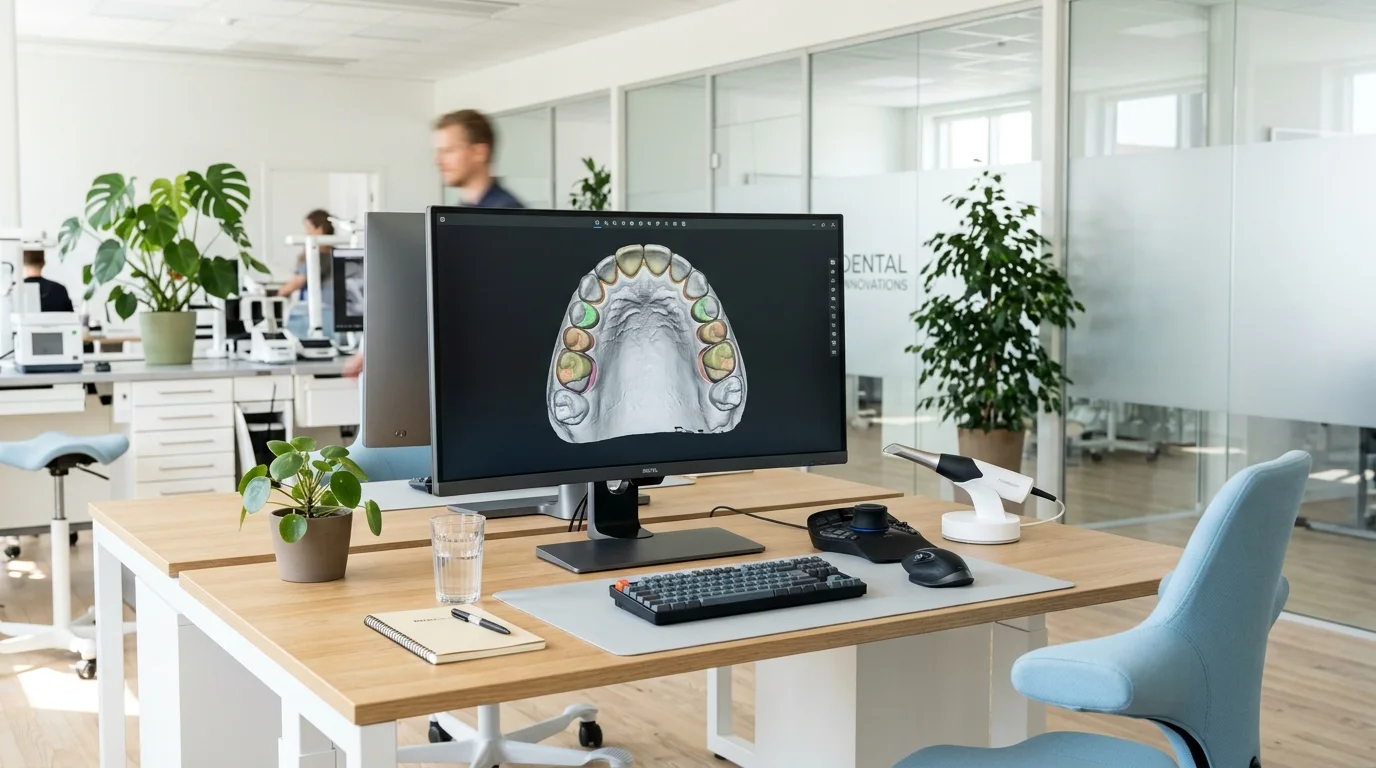
Intraoral scanners can only triangulate surfaces that are optically unobstructed. Crevicular fluid or collapsed gingiva obscures the chamfer in Exocad. Through our cloud STL intake workflow, our CAD engineers verify finish-line continuity within 15 minutes while your patient is still seated.
2. Double-Cord Deflection & Glare Control
- Two-Cord Protocol: Leave a fine #000 cord at the sulcus base and remove the top #0 cord immediately prior to scanning for 0.4 mm horizontal tissue clearance.
- Eliminate Saliva Pooling: Specular glare from moisture creates mesh spikes along the margin; air-dry gently before capturing the prep pass.
3. Selective HD Prep Mode & Bilateral Bite Scans
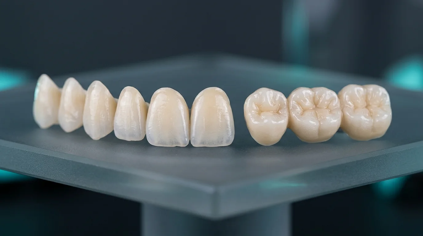
Activating high-density mesh capture selectively on prepared abutments delivers sub-15-micron accuracy on multilayer zirconia crowns and bridges. Always capture left and right buccal bite registrations with the patient seated upright.
DIGITAL WORKFLOW & FEE SCHEDULE
Upgrade your clinic’s digital prosthetic workflow
Pair your 3Shape, Medit Link, or iTero scanner and request our clinic fee schedule.