Chairside Bite Registration for Milled Digital Michigan Occlusal Splints
30-SECOND CLINICAL TAKEAWAYS
5 min read
- Scanning directly at the intended 2–2.5 mm vertical opening eliminates hinge-axis rotation errors.
- 5-axis milled cross-linked PMMA discs contain zero residual monomer and resist staining or fracture.
- Balanced centric stops with smooth canine disclusion protect new ceramic rehabilitations.
1. Why Scan at the Intended Vertical Opening?
Opening the bite virtually from MIP in CAD assumes an average hinge axis, often causing posterior premature contacts. Capturing your buccal bite scan using a leaf gauge or anterior deprogrammer at 2.0–2.5 mm posterior clearance ensures drop-in accuracy on milled Michigan occlusal splints.
2. Industrial Milled PMMA vs. Thermoformed Guards
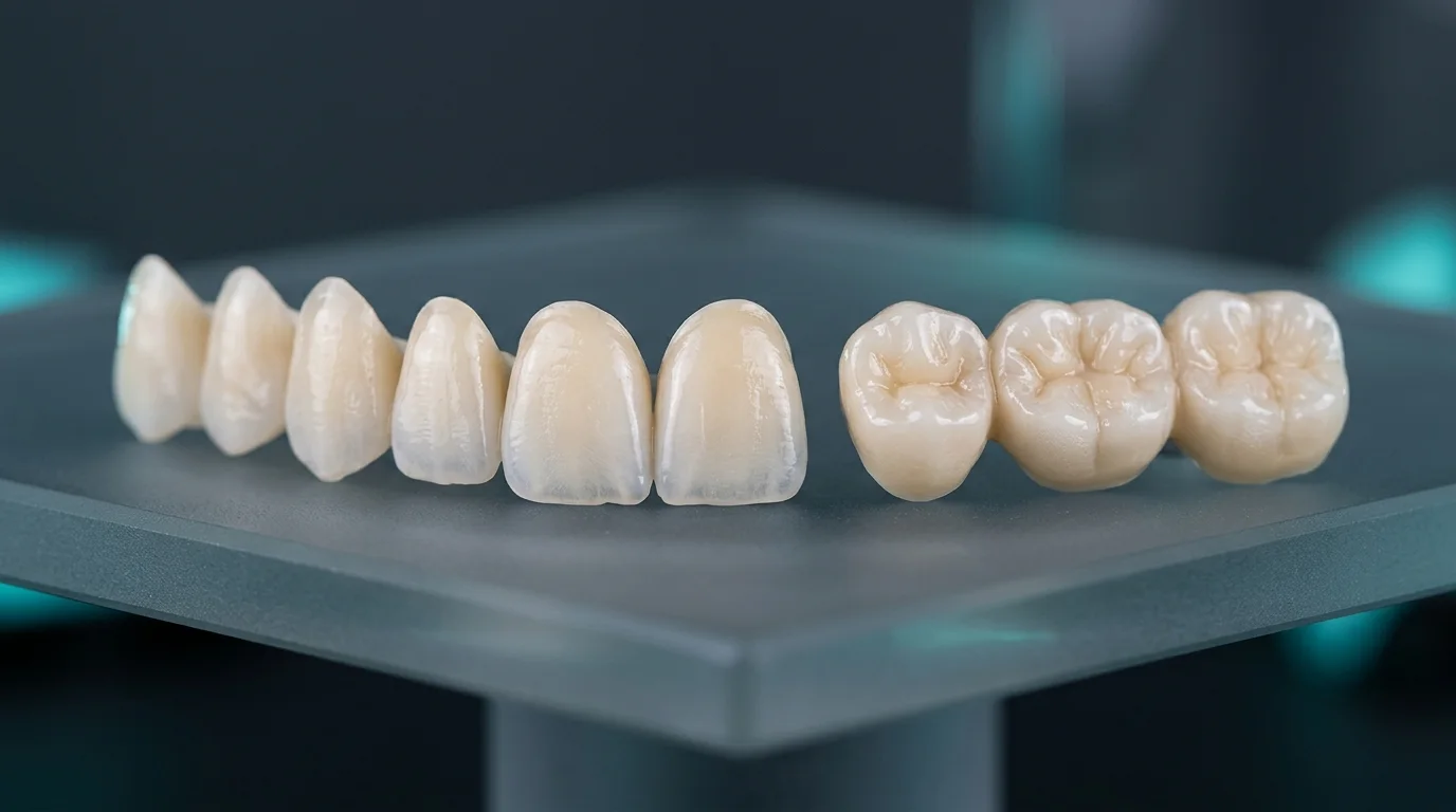
Machined from pre-polymerized optical-grade PMMA pucks, rigid stabilization splints provide smooth canine guidance that protects full-mouth zirconia and IPS e.max rehabilitations from nocturnal bruxism.
DIGITAL WORKFLOW & FEE SCHEDULE
Upgrade your clinic’s digital prosthetic workflow
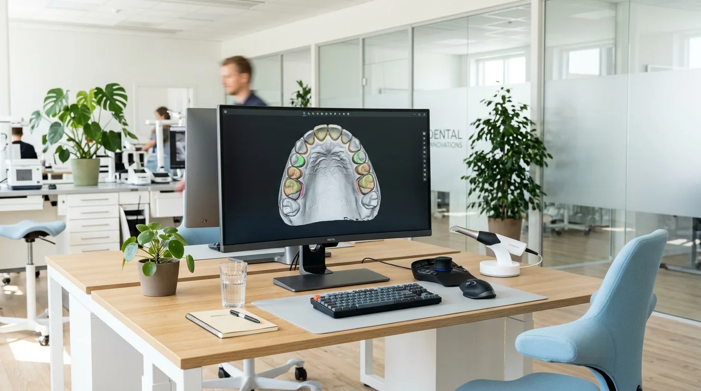
Pair your 3Shape, Medit Link, or iTero scanner and request our clinic fee schedule.